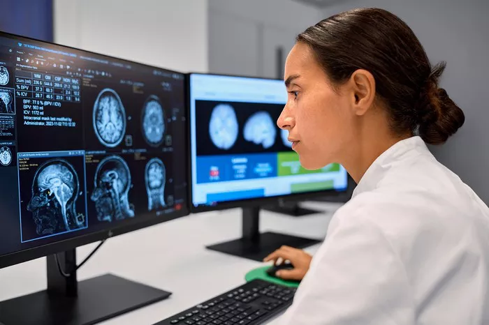
A cutting-edge machine learning model has proven more effective than existing techniques in harmonizing MRI brain volumetric data from different scanners while preserving Alzheimer’s disease markers, according to a study published in Radiology: Artificial Intelligence.
Quantitative markers like brain volume obtained from structural MRI scans are instrumental in diagnosing Alzheimer’s disease and distinguishing it from other dementias. Yet, differences in scanner hardware and imaging protocols have long plagued the reliability and reproducibility of automated MRI analyses—posing a major barrier to real-world clinical adoption.
Tackling MRI Scanner Variability with AI
To address this challenge, researchers led by Damiano Archetti, MSc, from IRCCS Fatebenefratelli in Italy, and Vikram Venkatrahavan, PhD, from Alzheimer Center Amsterdam, developed a new AI-based solution called the multiclass Neuroharmony model.
Unlike traditional harmonization techniques that often focus solely on healthy individuals, the multiclass version of Neuroharmony integrates data from both cognitively impaired and non-impaired individuals. The model uniquely incorporates cognitive status as a covariate, enabling it to preserve disease-related signatures while eliminating scanner-induced bias.
“Harmonization aims to remove protocol-related variability so the remaining data reflect true biological markers,” Archetti explained.
Large-Scale Validation Across Global Datasets
The researchers validated the model on a massive dataset of 20,864 participants from 11 cohorts, 43 MRI scanners, and three continents. Compared to other harmonization approaches—including a normative model trained solely on healthy individuals—the multiclass Neuroharmony model outperformed all alternatives, particularly on data from previously unseen scanners.
Crucially, the model was more effective in maintaining biologically meaningful differences between diagnostic groups, suggesting strong potential for clinical use.
“Our model showed better concordance across scanners while preserving key differences between healthy and pathological cases,” Archetti said.
Expert Opinions and Future Applications
In an accompanying editorial, Sven Haller, MD, a neuroradiologist and visiting professor in Sweden and China, praised the innovation, noting the frequent challenge of comparing baseline and follow-up scans acquired from different MR scanners due to facility changes or equipment upgrades.
“When subtle brain changes occur, it’s difficult to tell whether they reflect disease progression or scanner variability,” said Dr. Haller. “A harmonization tool that removes only the scanner-related differences would be very helpful.”
However, Dr. Haller also cautioned against tools that overcorrect and erase disease-related data.
“Completely separating disease-related from scanner-related variability remains an unsolved and highly complex challenge,” he added.
Archetti agreed, acknowledging that current AI models—including theirs—still have room for improvement.
Beyond Alzheimer’s: Toward Broader Neurological Applications
While this study focused specifically on the Alzheimer’s disease spectrum, researchers believe their approach could be extended to other neurological or psychiatric conditions, including those with major structural abnormalities such as brain lesions.
“Testing generalizability to other dementias or neurological diseases will be an important next step,” Archetti noted. “If the need for model adjustments arises, we’re ready to undertake that challenge.”
The multiclass Neuroharmony model represents a major stride toward trustworthy, cross-scanner MRI harmonization, with the potential to improve diagnostic consistency across imaging centers worldwide. As AI-driven medical tools mature, efforts like these bring us closer to more accurate and scalable brain health diagnostics.
Related topics:

